
Antibody and Dye Based Assays


Biotium Chromogranin A(CGA/414), CF488A conjugate, 0.1mg/mL
Chromogranin A is present in neuroendocrine cells throughout the body, including the neuroendocrine cells of the large and small intestine, adrenal medulla and pancreatic islets. It is an excellent marker for carcinoid tumors, pheochromocytomas, paragangliomas, and other neuroendocrine tumors. Co-expression of chromogranin A and neuron specific enolase (NSE) is common in neuroendocrine neoplasms. Reportedly, co-expression of certain keratins and chromogranin indicates neuroendocrine lineage. The presence of strong anti-chromogranin staining and absence of anti-keratin staining should raise the possibility of paraganglioma. The co-expression of chromogranin and NSE is typical of neuroendocrine neoplasms. Most pituitary adenomas and prolactinomas readily express chromogranin.Primary antibodies are available purified, or with a selection of fluorescent CF Dyes and other labels. CF Dyes offer exceptional brightness and photostability. Note: Conjugates of blue fluorescent dyes

Biotium CD3e (T-Cell Marker)(OKT3), CF488A conjugate, 0.1mg/mL
Recognizes the epsilon-chain of CD3, which consists of five different polypeptide chains (designated as gamma, delta, epsilon, zeta, and eta) with MW ranging from 16-28 kDa. CD3, also known as T3, is a member of the immunoglobulin superfamily that plays a role in antigen recognition, signal transduction, and T cell activation. It is found on all mature T-lymphocytes, NK, T-cells, and some thymocytes, and is expressed, typically at high levels, on peripheral T cells and majority of T cell neoplasms. Thymocytes express CD3 at different level on the cell surface in the course of differentiation and CD3 is predominantly intracytoplasmic in cortical thymus.Anti-CD3 clone OKT3 has been reported to have potent immunosuppressive properties in vivo and has been proven effective in the treatment of renal, heart and liver allograft rejection. In the crystal structure of the antibody-receptor complex, clone OKT3 was found to bind a conformational epitope on CD3e dominated by a linear
Biotium CD56 / NCAM(NCAM1/795), CF488A conjugate, 0.1mg/mL
This MAb reacts with an extracellular domain (close to transmembrane) of CD56/NCAM. Three isoforms of neural cell adhesion molecule (NCAM) are produced by differential splicing of the RNA transcript from a single gene. The 135 kDa isoform is the basic molecule, which is glycosylated or sialylated to produce the mature species. Anti-CD56 recognizes two proteins of the neural cell adhesion molecule, the basic molecule expressed on most neuroectodermally derived tissues and neoplasms (e.g. retinoblastoma, medulloblastomas, astrocytomas, neuroblastomas, and small cell carcinomas). It is also expressed on some mesodermally derived tumors (rhabdomyosarcoma). Anti-CD56 plays an important role in the diagnosis of nodal and nasal NK/T-cell lymphomas.Primary antibodies are available purified, or with a selection of fluorescent CF Dyes and other labels. CF Dyes offer exceptional brightness and photostability. Note: Conjugates of blue fluorescent dyes like CF405S and CF405M are not
Biotium Histiocytoma Marker(D11), CF488A conjugate, 0.1mg/mL
In Western blotting, this antibody detects an antigen of 125 kDa in human liver and 135 kDa in tumors of histiocytic origin. Comparative study of this MAb and a standard CD68 MAb showed that their antigens are different. Its antigen in all macrophage types studied is located on the plasma membrane and within cytoplasmic structures including lysosomes. This MAb shows a restricted reactivity to cells of the monocyte/macrophage system. It specifically reacts with blood monocytes and stains resident macrophages in a wide variety of human tissues. This MAb does not stain antigen-presenting cells, e.g., Langerhans cells. Reportedly, its reactivity is restricted to histiocytes and macrophages.Primary antibodies are available purified, or with a selection of fluorescent CF Dyes and other labels. CF Dyes offer exceptional brightness and photostability. Note: Conjugates of blue fluorescent dyes like CF405S and CF405M are not recommended for detecting low abundance targets, because
Biotium Histiocytoma Marker(D11), CF488A conjugate, 0.1mg/mL
In Western blotting, this antibody detects an antigen of 125 kDa in human liver and 135 kDa in tumors of histiocytic origin. Comparative study of this MAb and a standard CD68 MAb showed that their antigens are different. Its antigen in all macrophage types studied is located on the plasma membrane and within cytoplasmic structures including lysosomes. This MAb shows a restricted reactivity to cells of the monocyte/macrophage system. It specifically reacts with blood monocytes and stains resident macrophages in a wide variety of human tissues. This MAb does not stain antigen-presenting cells, e.g., Langerhans cells. Reportedly, its reactivity is restricted to histiocytes and macrophages.Primary antibodies are available purified, or with a selection of fluorescent CF Dyes and other labels. CF Dyes offer exceptional brightness and photostability. Note: Conjugates of blue fluorescent dyes like CF405S and CF405M are not recommended for detecting low abundance targets, because
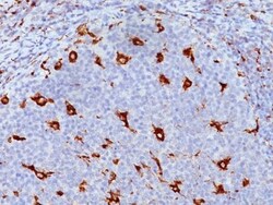
Biotium CD68(KP1), CF555 conjugate, 0.1mg/mL
This antibody recognizes a glycoprotein of 110 kDa, which is identified as CD68. It is important for identifying macrophages in tissue sections. It stains macrophages in a wide variety of human tissues, including Kupffer cells and macrophages in the red pulp of the spleen, in lamina propria of the gut, in lung alveoli, and in bone marrow. It reacts with myeloid precursors and peripheral blood granulocytes. It also reacts with plasmacytoid T cells, which are supposed to be of monocyte/macrophage origin. It shows strong granular cytoplasmic staining of chronic and acute myeloid leukemia and also reacts with rare cases of true histiocytic neoplasia. Lymphomas are negative or show few granules.Primary antibodies are available purified, or with a selection of fluorescent CF Dyes and other labels. CF Dyes offer exceptional brightness and photostability. Note: Conjugates of blue fluorescent dyes like CF405S and CF405M are not recommended for detecting low abundance targets,
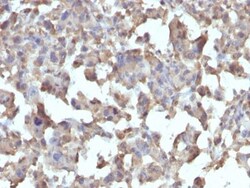
Biotium CD284 / TLR4(TLR4/230), CF488A conjugate, 0.1mg/mL
This MAb reacts with human Toll-like receptor 2 (TLR4). It is a member of the Toll-like receptor (TLR) family, which plays a fundamental role in pathogen recognition and activation of innate immunity. TLRs are highly conserved from Drosophila to humans and share structural and functional similarities. They recognize pathogen-associated molecular patterns that are expressed on infectious agents, and mediate the production of cytokines necessary for the development of effective immunity. The various TLRs exhibit different patterns of expression. This receptor has been implicated in signal transduction events induced by lipopolysaccharide (LPS) found in most gram-negative bacteria. Mutations in this gene have been associated with differences in LPS responsiveness. Multiple transcript variants encoding different isoforms have been found for this gene.Primary antibodies are available purified, or with a selection of fluorescent CF Dyes and other labels. CF Dyes offer exceptional
Biotium CD7(B-F12), CF488A conjugate, 0.1mg/mL
Recognizes a protein of 40 kDa, identified as CD7 (also known as gp40, Leu9). CD7 is a member of the immunoglobulin gene superfamily. Its N-terminal amino acids 1-107 are highly homologous to Ig kappa-L chains whereas the carboxyl-terminal region of the extracellular domain is proline-rich and has been postulated to form a stalk from which the Ig domain projects. CD7 is expressed on the majority of immature and mature T-lymphocytes, and T cell leukemia. It is also found on natural killer cells, a small subpopulation of normal B cells and on malignant B cells. Cross-linking surface CD7 positively modulates T cell and NK cell activity as measured by calcium fluxes, expression of adhesion molecules, cytokine secretion and proliferation. CD7 associates directly with phosphoinositol 3'-kinase. CD7 ligation induces production of D-3 phosphoinositides and tyrosine phosphorylation.Primary antibodies are available purified, or with a selection of fluorescent CF Dyes and other labels.
Biotium Cytokeratin 7(KRT7/1198), CF488A conjugate, 0.1mg/mL
This antibody recognizes an intermediate filament protein (IFP) of 55 kDa, which is identified as cytokeratin 7. This MAb is highly specific to cytokeratin 7 and shows no cross-reaction with other IFPs. Cytokeratin 7 is a basic cytokeratin, which is found in most glandular and transitional epithelia but not in the stratified squamous epithelia. Keratin 7 is expressed in the epithelial cells of ovary, lung, and breast but not of colon, prostate, or gastrointestinal tract. This MAb is highly useful in distinguishing ovarian carcinomas (keratin 7 ) from colon carcinomas (keratin 7-).Primary antibodies are available purified, or with a selection of fluorescent CF Dyes and other labels. CF Dyes offer exceptional brightness and photostability. Note: Conjugates of blue fluorescent dyes like CF405S and CF405M are not recommended for detecting low abundance targets, because blue dyes have lower fluorescence and can give higher non-specific background than other dye colors.
Biotium CD56 / NCAM(123C3.D5), CF488A conjugate, 0.1mg/mL
This MAb reacts with an extracellular domain (close to transmembrane) of CD56/NCAM. Three isoforms of neural cell adhesion molecule (NCAM) are produced by differential splicing of the RNA transcript from a single gene. The 135 kDa isoform is the basic molecule, which is glycosylated or sialylated to produce the mature species. Anti-CD56 recognizes two proteins of the neural cell adhesion molecule, the basic molecule expressed on most neuroectodermally derived tissues and neoplasms (e.g. retinoblastoma, medulloblastomas, astrocytomas, neuroblastomas, and small cell carcinomas). It is also expressed on some mesodermally derived tumors (rhabdomyosarcoma). Anti-CD56 plays an important role in the diagnosis of nodal and nasal NK/T-cell lymphomas.Primary antibodies are available purified, or with a selection of fluorescent CF Dyes and other labels. CF Dyes offer exceptional brightness and photostability. Note: Conjugates of blue fluorescent dyes like CF405S and CF405M are
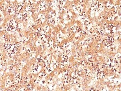
Biotium AFP (Alpha Fetoprotein)(C3), CF488A conjugate, 0.1mg/mL
Alpha-fetoprotein (AFP) is an oncofetal glycoprotein with a single chain of 70 kDa. This MAb is highly specific to AFP and shows no cross-reaction with other oncofetal antigens or serum albumin. AFP is normally synthesized in the liver, intestinal tract, and yolk sac of the fetus. Antibody to AFP has been shown to be useful in detecting hepatocellular carcinomas (HCC) and germ cell neoplasms, especially yolk sac tumors.Primary antibodies are available purified, or with a selection of fluorescent CF Dyes and other labels. CF Dyes offer exceptional brightness and photostability. Note: Conjugates of blue fluorescent dyes like CF405S and CF405M are not recommended for detecting low abundance targets, because blue dyes have lower fluorescence and can give higher non-specific background than other dye colors.
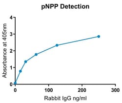
Abcam Alkaline Phosphatase Conjugation Kit (Animal Free) - Lightning-Link, 100UG
Alkaline Phosphatase conjugation / labeling in < 4 hrs with 30 secs hands-on time using Alkaline Phosphatase Conjugation Kit (Animal Free) - Lightning-Link ab269901. 100% antibody recovery.
The product is subject to the following: Abcam Restricted Use Statement


Medchemexpress LLC (E)-9-amino-10-methyl-2-(naphthalen-1-ylmethylene)-1-oxo-1,2,3,4-tetrahydroacridin-10-ium iodide | 2252162-81-3 | 98.1% | C25H21IN2O | 10MG

Aβ/tau aggregation-IN-1 is a small-molecule research inhibitor that reduces aggregation of amyloid-β (Aβ1-42) and tau by preventing β-sheet formation and disrupting aggregate interactions. It demonstrates activity in cell-based tau aggregation assays and has documented physicochemical and storage properties for preclinical use.
- Inhibits aggregation of Aβ1-42 and tau, reducing β-sheet formation.
- Reported KD values ≈160 μM for Aβ1-42 and ≈337 μM for tau.
- Reduces tau aggregation foci at 1 μM in HEK-293T cellular assays.
- High purity (98.1%) suitable for research applications.
- Soluble in DMSO (~33.3 mg/mL); in vivo formulation examples provided.
- Solid storage at 4°C; in solvent store at -80°C for long-term stability.
- For research use only; not for human use.
Biotium bcl-2(8C8), CF488A conjugate, 0.1mg/mL
This antibody recognizes a protein of 25-26 kDa, identified as Bcl-2. This antibody shows no cross-reaction with Bcl-x or Bax proteins. Bcl-2 suppresses apoptosis in a variety of cell systems including factor-dependent lymphohematopoietic and neural cells. It regulates cell death by controlling the mitochondrial membrane permeability. Bcl-2 appears to function in a feedback loop system with caspases. It inhibits caspase activity either by preventing the release of cytochrome c from the mitochondria and/or by binding to the apoptosis-activating factor (APAF-1). Expression of Bcl-2 inhibits programmed cell death (apoptosis). In most follicular lymphomas, neoplastic germinal centers express high levels of Bcl-2 protein, whereas the normal or hyperplastic germinal centers are negative. Consequently, this antibody is valuable when distinguishing between reactive and neoplastic follicular proliferation in lymph node biopsies. It may also be used in distinguishing between those foll